

Cryo-electron microscopy (Cryo-EM) has essentially reworked our understanding of the molecular equipment that governs life. For many years, structural biology was usually restricted by the “crystallization bottleneck” of X-ray diffraction. Cryo-EM modified the paradigm, permitting scientists to look into the microscopic world with unprecedented readability and element.
With its origins rooted within the pioneering work of Nobel laureates Jacques Dubochet, Joachim Frank, and Richard Henderson, Cryo-EM has advanced from a distinct segment specialty—as soon as jokingly known as “blobology”—right into a mainstream powerhouse. Immediately, it’s able to revealing the hidden constructions of proteins, viruses, and mobile elements at atomic decision.
What’s Cryo-EM?
Cryo-EM is an umbrella time period for a set of superior imaging strategies used to elucidate the three-dimensional (3D) construction of organic molecules and complexes of their native, hydrated state.
On this approach, the pattern is quickly frozen to temperatures beneath -150 °C, trapping it in vitreous ice. By imaging these frozen samples from totally different angles utilizing a transmission electron microscope (cryo-TEM), researchers seize “snapshots” of molecules as they exist in resolution. Superior computational algorithms then digitally recombine these 2D snapshots right into a high-fidelity 3D reconstruction.
The Core Precept: Vitrification
The organic relevance of Cryo-EM hinges on Vitrification. In typical electron microscopy, cooling water normally results in the formation of crystalline ice, which expands and destroys delicate protein constructions. Moreover, crystalline ice causes sturdy electron diffraction that dramatically reduces decision.
Vitrification entails flash-freezing the aqueous resolution so rapidly (inside milliseconds) that the water molecules shouldn’t have time to crystallize. As an alternative, they type a vitreous strong—a glass-like state that’s clear to electrons. This course of supplies a novel benefit: it preserves the specimen in its near-native state, offering physiologically related observations that strategies requiring harsh chemical therapies, staining, or dehydration can not match.
Historic Growth and the Nobel Path
The event of Cryo-EM was a direct response to the restrictions of typical EM, particularly pattern destruction brought on by high-energy electron beams and artifacts derived from chemical fixation.
Seventies: Richard Henderson and Nigel Unwin produced the primary 3D construction of a organic specimen (bacteriorhodopsin) utilizing EM.
Eighties: Jacques Dubochet developed the methodology for vitrifying water, permitting organic samples to be ready for vacuum situations with out shedding their construction.
Nineteen Nineties: Joachim Frank developed the computational methods to course of 2D pictures of randomly oriented particles into 3D constructions.
2013–Current: The “Decision Revolution” started with the event of Direct Electron Detectors and improved software program, resulting in the 2017 Nobel Prize in Chemistry.
Comparability: Standard EM vs. Cryo-EM
FeatureConventional EMCryo-EMPattern PrepFixation, staining, dehydrationVitrification (Flash-freezing)IntegrityExcessive threat of artifactsNative construction preservedDecisionNanometer (nm)Close to-atomic (Å)Pattern QuantityBigger quantities requiredMinimal (µg/mL)
The Three Pillars of Cryo-EM Methods
Cryo-EM is just not a single technique however a set of analytical instruments tailor-made to the character of the pattern.
1. Single Particle Evaluation (SPA)
The most well-liked Cryo-EM approach and the first different to X-ray crystallography.
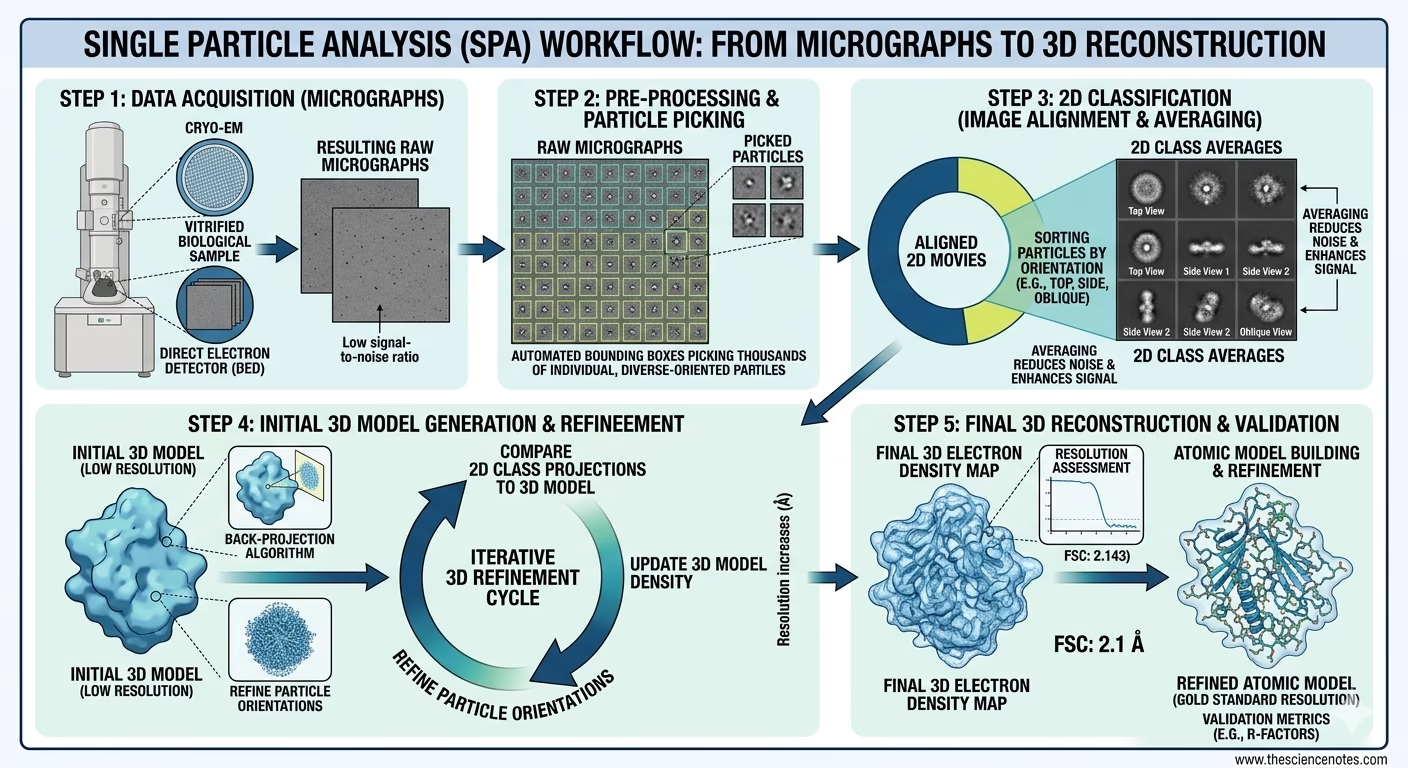
The Course of: Purified proteins are vitrified, leading to 1000’s of molecules oriented randomly inside the ice.
The Objective: The TEM collects 2D snapshots of each potential orientation. These are then digitally aligned, labeled, and averaged right into a 3D reconstruction.
Key Achievement: SPA has just lately resolved constructions just like the human GABA-A receptor at 1.7 Å, revealing binding pockets for small molecules like histamine.
2. Cryo-Electron Tomography (Cryo-ET)
Whereas SPA appears to be like at “purified components,” Cryo-ET appears to be like on the “entire machine” contained in the cell.
The Course of: Used for entire cells or tissues. Since cells are thick, researchers use Targeted Ion Beam (FIB) milling to shave away layers, creating a skinny “window” or lamella.
The Objective: The pattern is tilted at varied angles ($+/- 60) to take a collection of 2D pictures (a tomographic tilt collection) which might be reconstructed right into a 3D dataset referred to as a tomogram.
3. Microcrystal Electron Diffraction (MicroED)
MicroED is the “bridge” between crystallography and EM.
The Course of: It makes use of electron diffraction relatively than imaging.
The Benefit: Electrons work together with matter 10^4 to 10^6 instances extra strongly than X-rays. This permits atomic particulars to be extracted from nanocrystals (<200 nm) which might be too small for conventional X-ray diffraction.
How Cryo-EM Works: The Technical Workflow
The fashionable Cryo-EM workflow has advanced from a handbook, labor-intensive course of into an more and more automated pipeline. The transition from a purified pattern to a high-resolution 3D density map entails 4 vital phases:
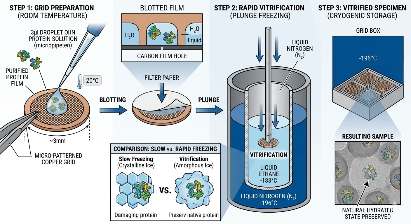
1. Pattern Preparation and Grid Loading
The method begins with a purified macromolecule suspended in an optimized aqueous buffer. A tiny quantity (usually 3–5 microliters) is utilized to a specialised EM grid—normally a 3mm copper mesh coated with a skinny, holey carbon or gold movie.
Vital Issue: The focus have to be exactly tuned to make sure that particles are densely packed inside the grid holes however don’t overlap, which might complicate the later phases of particle choosing.
2. Vitrification: The Artwork of Flash-Freezing
The grid is mechanically “blotted” with filter paper to take away extra liquid, leaving a skinny movie of water only some hundred nanometers thick. The grid is then quickly plunged right into a major coolant, usually liquid ethane (maintained at approx. -183°C).
3. Excessive-Decision Knowledge Acquisition (Imaging)
The vitrified grids are transferred into the high-vacuum setting of a Cryo-TEM (Transmission Electron Microscope) or Cryo-SEM (Scanning Electron Microscope).
Cryo-TEM: The electron beam passes via the pattern to disclose inside atomic constructions. That is the first instrument for Single Particle Evaluation.
Direct Electron Detectors (DEDs): Fashionable microscopes use CMOS-based DEDs that act like high-speed cameras, capturing “films” relatively than static pictures. This permits the software program to trace and proper for the motion of the pattern throughout the publicity.
4. Computational Picture Processing
As soon as terabytes of uncooked “films” are collected, they bear rigorous digital evaluation:
Movement Correction: The software program aligns particular person film frames to repair “beam-induced movement”—the slight motion triggered when electrons strike the frozen pattern.
CTF Estimation: Calculating the Distinction Switch Perform to appropriate for lens aberrations and defocus, guaranteeing the ultimate map is sharp and correct.
Particle Choosing: Automated algorithms (usually powered by Deep Studying/AI) establish and extract lots of of 1000’s of particular person protein projections from the micrographs.
2D Classification: These projections are sorted into teams based mostly on their orientation. “Dangerous” particles (broken proteins or contaminants) are discarded right here.
3D Refinement: Via iterative back-projection algorithms, the 2D courses are merged right into a 3D density map. This map is refined till the decision permits for the constructing of an atomic mannequin.
Arithmetic in Knowledge Evaluation
To attain atomic decision, Cryo-EM depends on advanced sign processing to beat low distinction.
The Distinction Switch Perform (CTF)
The microscope optics introduce part shifts described by the CTF:
CTF(okay) = A(okay) sin [ π Δz λ k² + ½ π Cs λ³ k⁴ ]
The place:
Fourier Shell Correlation (FSC)
The standard of a 3D map is decided by the FSC. The info is break up into two impartial halves; the decision is outlined on the level (usually the 0.143 threshold) the place the correlation between the 2 maps drops considerably.
Strengths, Limitations, and Comparisons
Cryo-EM serves as a robust, complementary approach to different structural biology strategies corresponding to Nuclear Magnetic Resonance (NMR) and X-ray Crystallography.
Strengths
No Crystallization Required: Eliminates the only greatest bottleneck in structural biology, notably for big complexes and membrane proteins.
Visualizes Giant, Versatile Complexes: Excels at resolving huge molecular machines (like ribosomes) which might be too advanced for NMR.
Captures Conformational Heterogeneity: In contrast to the static “common” of a crystal, Cryo-EM can seize a protein “in movement,” displaying a number of practical states in a single pattern.
Limitations
The Small Protein Barrier: Molecules smaller than < 50 kDa are presently tough to picture and align as a result of their low signal-to-noise ratio.
Excessive Monetary Barrier: A top-tier setup represents a major funding, with tools prices starting from $5 to $7 million USD, plus excessive annual upkeep.
Huge Knowledge Demand: A single session can generate a number of terabytes of uncooked information, necessitating sturdy GPU-based computational clusters for processing and storage.
Purposes of Cryo-EM
Structural Biology: Understanding protein features and mechanisms of membrane proteins (GPCRs).
Virology: Aiding vaccine improvement by visualizing viral capsids and spike proteins (e.g., SARS-CoV-2).
Drug Discovery: Rational drug design for ion channels and huge macromolecular assemblies.
Neurobiology: Learning synaptic vesicles and protein aggregates in neurodegenerative ailments like Alzheimer’s.
Biochemistry: Mapping the equipment of DNA replication, transcription, and translation.
Conclusion
Cryo-electron microscopy has moved from a specialty instrument to a transformative expertise. By enabling the visualization of organic samples of their near-native state and at excessive decision, it supplies unprecedented insights into the constructions and features of life’s numerous biomolecular techniques. Whether or not via Single Particle Evaluation, Cryo-Tomography, or MicroED, it permits us to research proteins in all their advanced, heterogeneous glory.
X-ray Crystallography: Definition, Precept, Steps, Knowledge Evaluation, Purposes, and Limitations