

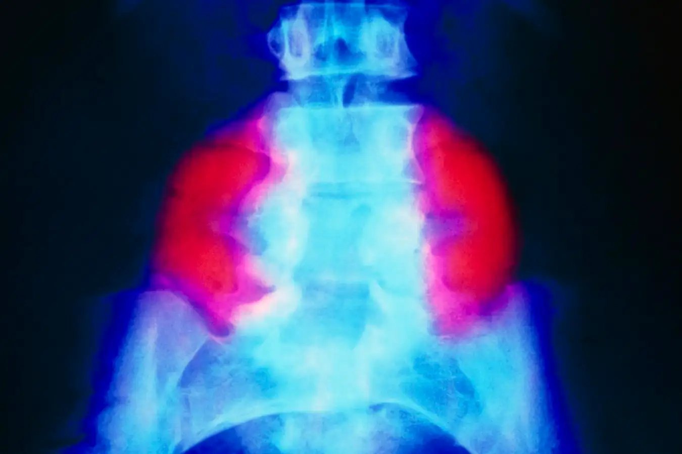
A false-colour X-ray exhibiting a big neural tube defect (crimson) on each side of the decrease again in somebody with spina bifida
SCIENCE PHOTO LIBRARY
A patch made from stem cells from donor placentas has been used to deal with fetuses within the womb with a extreme type of spina bifida as a part of a world-first trial. The novel method appears to have reversed a mind complication related to the congenital situation at the least as successfully because the go-to remedy, however is anticipated to allow extra youngsters to stroll over the long run.
The mom of one of many infants, who’s now 4 years previous, says she anticipated that her son Toby would require a wheelchair when he was identified with the situation within the womb. “However Toby is wholesome [and] has hit all of his milestones – he’s strolling, operating and leaping – and has no issues with bladder management, which is uncommon for individuals with the situation,” she says.
Spina bifida – which impacts about 1 in each 2800 births within the US yearly – happens when a child’s backbone and spinal twine don’t totally develop within the womb. In essentially the most extreme type of the situation, known as myelomeningocele, the spinal twine and its surrounding tissue protrude out of a niche within the vertebrae, which regularly impairs mobility and bowel and bladder management. The reason for spina bifida is unknown, however folic acid deficiency throughout being pregnant raises the danger.
One of many normal therapies includes surgical procedure within the womb that tucks the spinal twine and the encircling tissue again into the vertebrae, earlier than stitching up the pores and skin to type a decent seal. “However many youngsters nonetheless find yourself unable to stroll and there’s [usually] no enchancment in bowel or bladder management,” says Diana Farmer on the College of California, Davis.
This led Farmer and her colleagues to marvel if the addition of stem cells might assist by selling the expansion and restore of spinal tissue. To seek out out, they recruited six pregnant ladies carrying fetuses with myelomeningocele.
By about 24 weeks’ gestation, all of the fetuses had developed a typical complication known as hindbrain herniation, the place an excessive amount of fluid builds up within the cranium, pushing the underside of the mind, the cerebellum, via a gap on the cranium’s base. The usual surgical procedure typically helps to reverse hindbrain herniation, however many youngsters nonetheless have problems.
Within the newest trial, all of the fetuses underwent the usual surgical procedure but additionally obtained a patch, measuring a number of centimetres lengthy, that contained stem cells derived from donated placentas that have been embedded in a matrix of sticky proteins. Surgeons positioned this patch on the backbone earlier than the pores and skin was sewn round it. “The cells secrete their magic stem cell juice,” says Farmer.
At start, the surgical procedure web site had healed properly in the entire infants, with no indicators of irregular cell progress. “A key fear was that including stem cells in a fetus would make the cells develop like loopy, however we didn’t see that,” says Farmer. MRI scans of their brains additionally confirmed that the remedy utterly reversed hindbrain herniation.
“My private opinion is that this may enhance long-term outcomes in comparison with the usual method [based on evidence from animal studies],” says Panicos Shangaris at King’s School London.
The researchers hope to evaluate this in a trial the place 35 fetuses with myelomeningocele will obtain the stem-cell patch, and their outcomes will probably be in contrast towards a earlier research that used the standard surgical procedure, says Farmer.
However Shangaris says that a greater comparability, which is extra more likely to result in the remedy being permitted, could be to match the 2 approaches in a head-to-head trial that assesses their security and efficacy on fetuses who have been randomly assigned to every intervention.
Subjects: